Solution
v = u - gT
v = 0 at top most point.
T =
Velocity of the ball after (T - t) second
v = u - g(T - t)
= u - gT + gt = u - g + gt
v = gt
Distance covered during the last t seconds of its ascent is
s = (gt)t - =


| AgI | ⇌ | Ag+ | + | I- |
|---|---|---|---|---|
| s | s | s |


In the ABO blood group system, there are two main antigens (proteins on the surface of red blood cells) that are considered: antigen A and antigen B. An individual's blood type in this system is determined by the presence or absence of these antigens. Additionally, in the bloodstream, there will usually be antibodies against the antigens that the individual does not express. For example, someone with antigen A will typically have anti-B antibodies, and someone with antigen B will typically have anti-A antibodies. A person with neither antigen will have both types of antibodies, and a person with both antigens will have neither type of antibodies.
If both antigens are present (antigen A and antigen B) on the surface of the red blood cells but no antibodies against them are found in the plasma, this signifies that the blood group of the individual is AB. This is because the presence of both antigens means the immune system does not need to produce antibodies against them since the body recognizes both antigens as self.
Therefore, the correct answer is:
Option C: AB
The correct answer is Option B: muscle fibres found only in the ventricle wall.
Let's elaborate on the nature and function of the Bundle of His to clarify this answer. The Bundle of His, also known as the Atrioventricular Bundle (AV Bundle), is a crucial part of the electrical conduction system of the heart. It is not a network of nerve fibers but rather specialized muscle fibers.
The heart's electrical conduction system is responsible for coordinating the heartbeat, ensuring that blood is pumped efficiently throughout the body. The sequence of the conduction system goes as follows: from the sinoatrial (SA) node, located in the right atrium, the electrical impulse travels to the atrioventricular (AV) node. From the AV node, the impulse passes down through the Bundle of His, which then divides into the right and left bundle branches that run along the sides of the ventricular septum.
The Bundle of His is specifically located between the atria and the ventricles within the heart's septum—the wall dividing the right and left sides of the heart. Its role is critical for transmitting electrical impulses from the atria to the ventricles, a process essential for maintaining a synchronized heartbeat. After the impulse moves through the Bundle of His, it travels down through the right and left bundle branches and into the Purkinje fibers, spreading throughout the ventricles and causing them to contract.
Thus, the Bundle of His is comprised of specialized cardiac muscle fibers, not nerve fibers, and is found specifically in the heart's ventricular walls, bridging the electrical conduction between the atria and the ventricles. This corrects the common misconception that it might be composed of nerve fibers or distributed throughout the entire heart, including the atrial walls.
The most appropriate site for grafting an artificial pacemaker is at the site of the sinoatrial (SA) node, making option C the correct answer. Let's explore why that is the case by understanding the role of the SA node and the other options provided.
The SA node, located in the right atrium of the heart, acts as the natural pacemaker of the heart. It generates electrical impulses that initiate each heartbeat, regulating heart rate. These impulses travel across the atria, causing them to contract and pump blood into the ventricles. This is why, when the natural pacemaker fails to function properly, an artificial pacemaker is often installed to mimic the function of the SA node, ensuring the heart maintains a proper rhythm.
Now, let's briefly look at the other options:
Option A: Atrioventricular Bundle - Also known as the Bundle of His, this pathway transmits impulses from the atria to the ventricles. While it's crucial for the coordinated contraction of the ventricles, it is not the primary site for initiating the heart's rhythm.
Option B: Purkinje System - This network spreads throughout the ventricles and ensures efficient contraction of the ventricular muscle. It's essential for the final distribution of the electrical impulse but not the initiation of the heart rhythm.
Option D: Atrioventricular Node - Located at the junction of the atria and ventricles, the AV node delays the electrical impulse slightly before passing it to the ventricles. This delay allows the atria to finish contracting before the ventricles begin. While important for coordinating the timing of contractions, it is not the site where the heart's rhythm is generated.
Therefore, among the options provided, the sinoatrial node (Option C) is the most suitable site for grafting an artificial pacemaker, as it takes over the role of initiating and regulating the heart rhythm.
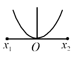
The correct option is Option D, which states that Fasciola hepatica (liver fluke) infects its intermediate host and primary host at the larval stages of miracidium and metacercaria respectively. Here's a brief overview of the life cycle of Fasciola hepatica to explain why:
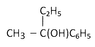
1. Miracidium: The life cycle begins when eggs released into the environment from an infected primary host (typically a sheep or a cow) hatch into miracidia upon contact with water. The miracidium is equipped to infect the intermediate host, which is a specific type of freshwater snail.
2. Redia & Cercaria: Inside the snail, the miracidium undergoes several developmental stages, initially transforming into sporocysts, then into rediae, and finally into cercariae. The redia stage is particularly noteworthy because within the snail, it will produce several generations, which increase the number of cercariae produced. The cercariae are then released from the snail into the water.
3. Metacercaria: Once free-swimming, the cercariae encyst as metacercariae on aquatic vegetation or other surfaces. This encysted stage is infectious to the primary host. When the primary host ingests these metacercariae-laden plants or water, the life cycle progresses.
4. Adult: Inside the primary host's intestines, the metacercariae excyst and migrate to the liver, where they mature into adult flukes, completing the cycle.
Therefore, the miracidium phase targets the intermediate snail host, initiating internal development, while the metacercaria phase represents the stage at which Fasciola hepatica is capable of infecting the primary host, marking the transition from external to internal parasitism in the latter.